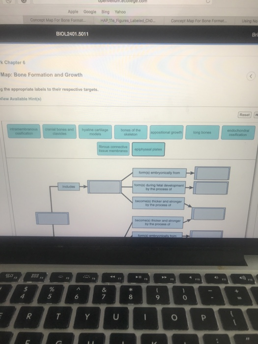
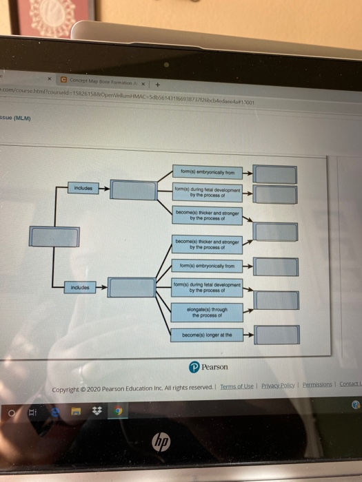
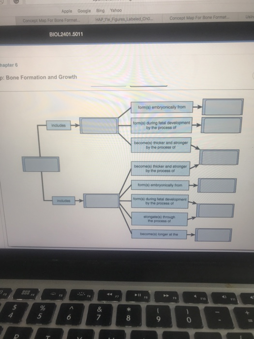
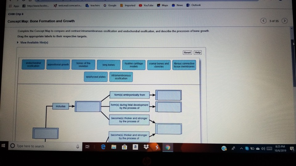
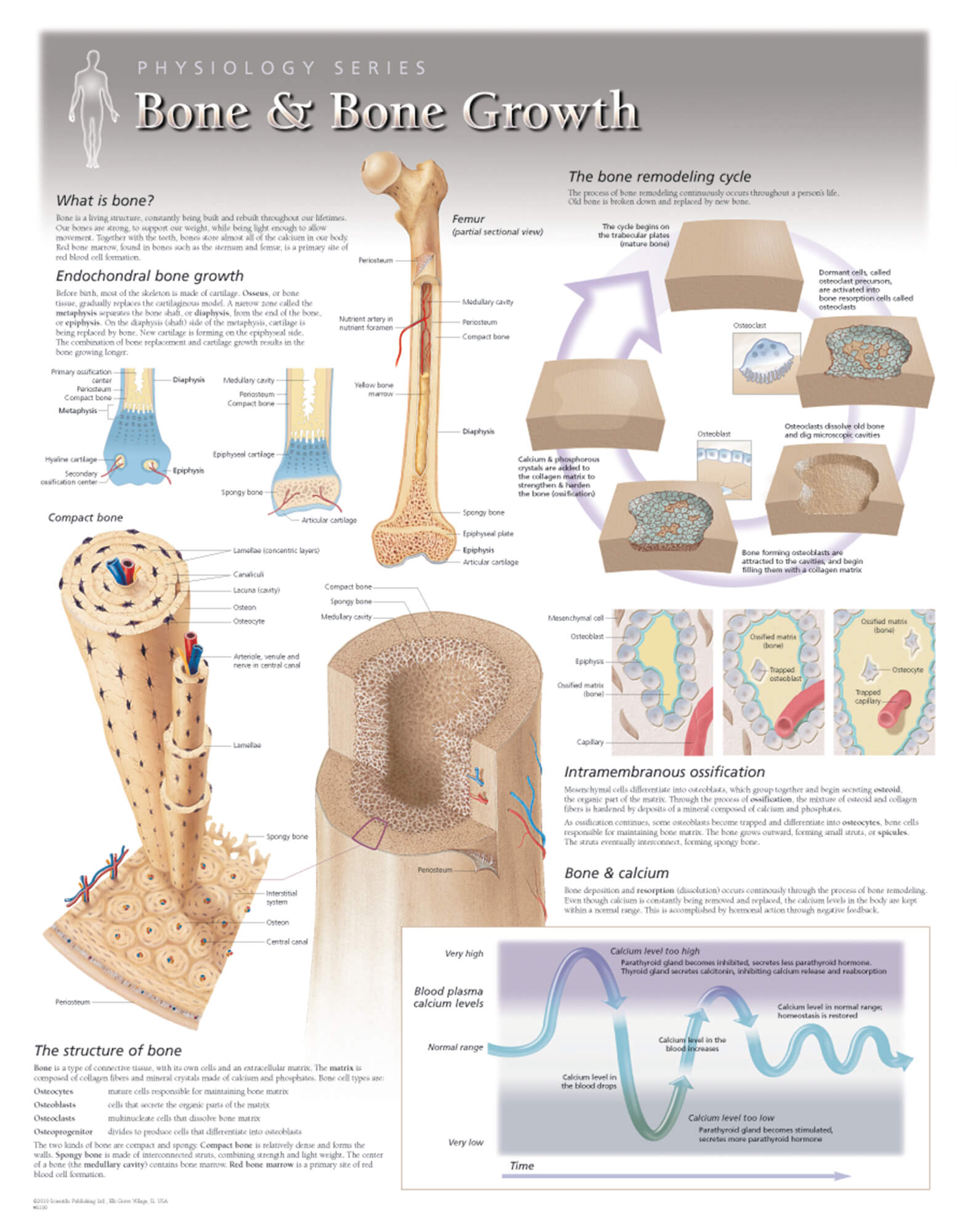
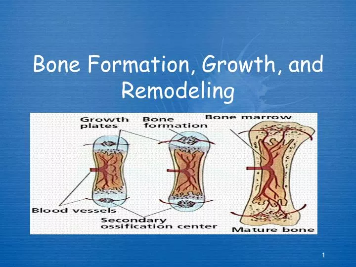
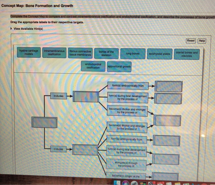
Endochondral ossification follows five steps.The process begins when mesenchymal cells in the embryonic skeleton gather together and begin to differentiate into specialized cells (figure 5.4.1.a 5.4.
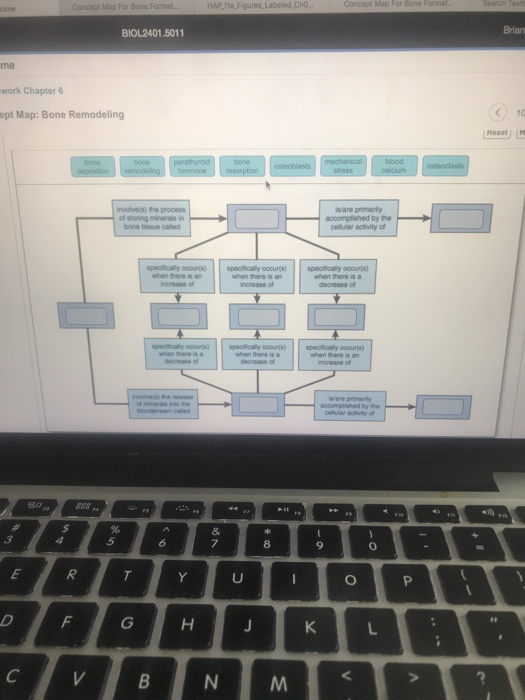
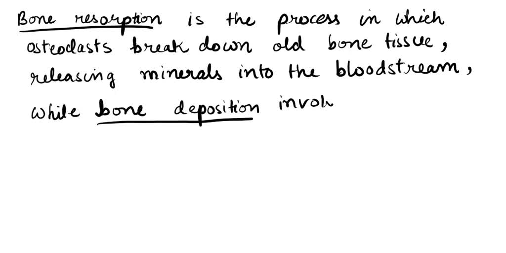
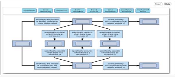
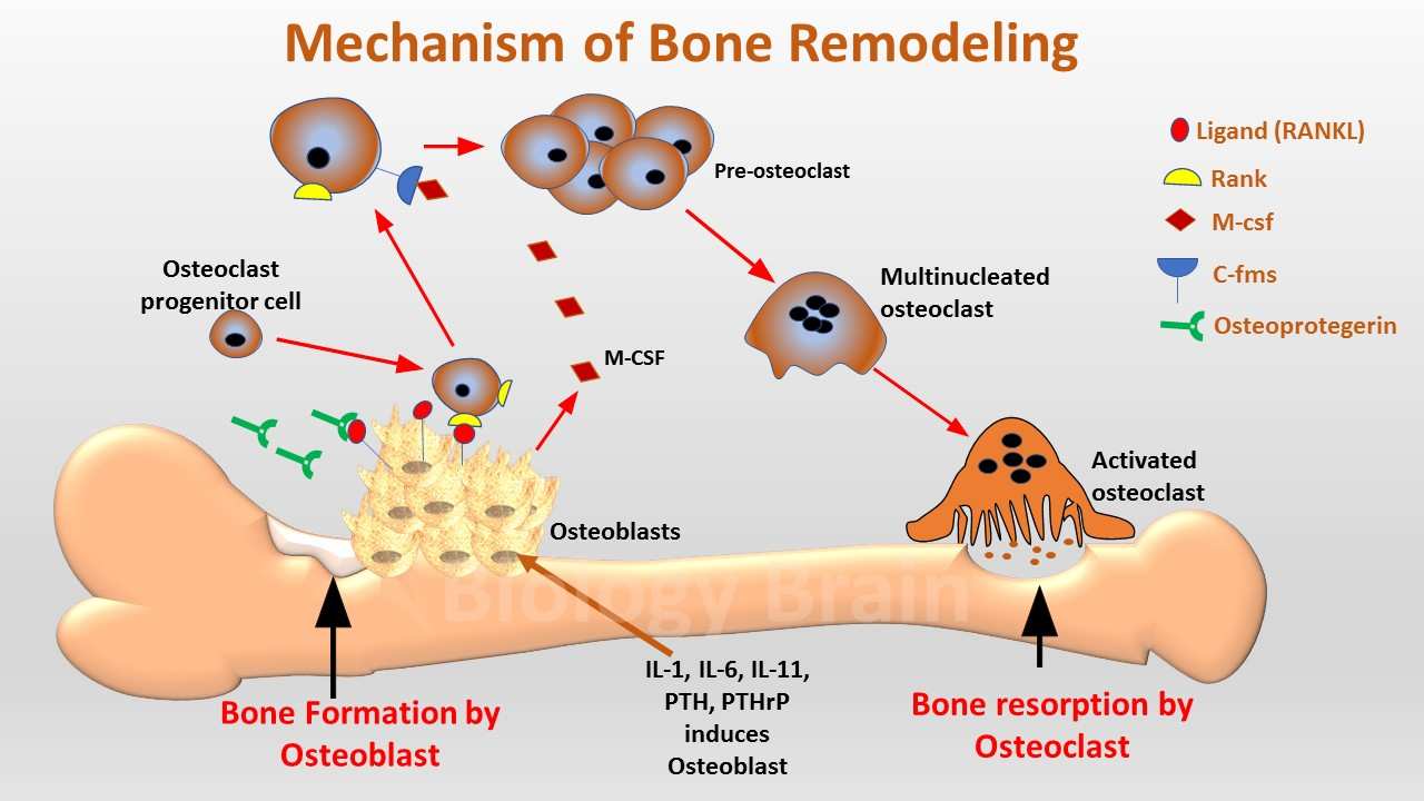
Mature cells in both live in lacunae, 3.On the contrary, endochondral ossification is dependent on a cartilage model.The balance between the two phases is crucial for sustaining bone mass and systemic mineral homeostasis.
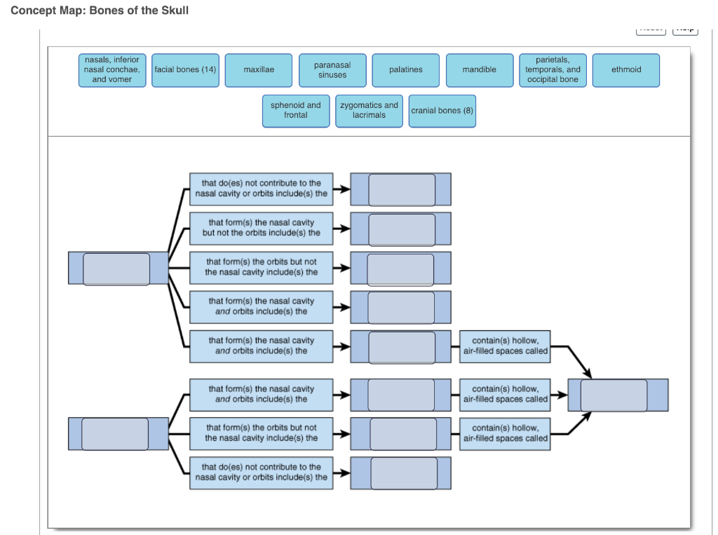
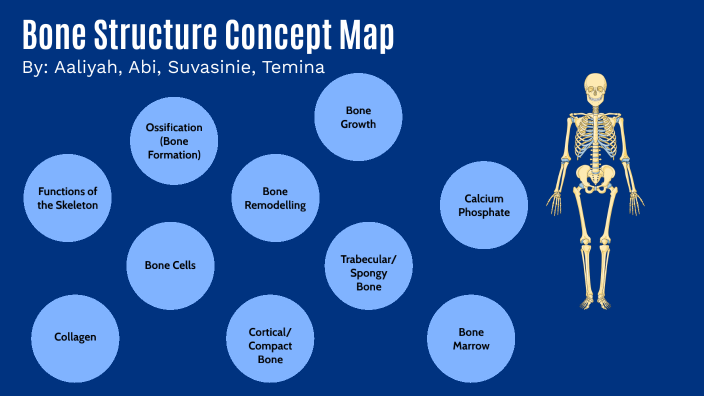
Long and short bones, such as the femur and phalanges, arise from a cartilage.Name 4 ways cartilage and bone are different.
In the early stages of embryonic development, the embryo's skeleton consists of fibrous membranes and hyaline cartilage.Of the articular and epiphyseal cartilage.From the genesis of embryonic development to the dynamic remodeling that sustains us throughout adulthood, this exploration illuminates the cellular, hormonal, and systemic factors that orchestrate the.
This model continues to grow as ossification takes place.This review highlights recent work on physiological bone remodeling and discusses our knowledge of how systemic and growth factors regulate this process.
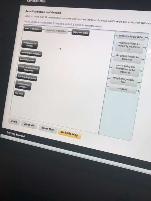
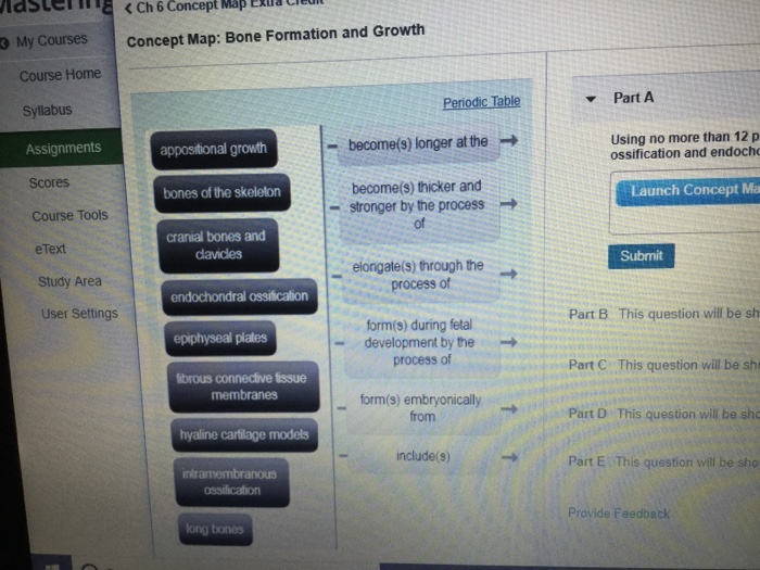
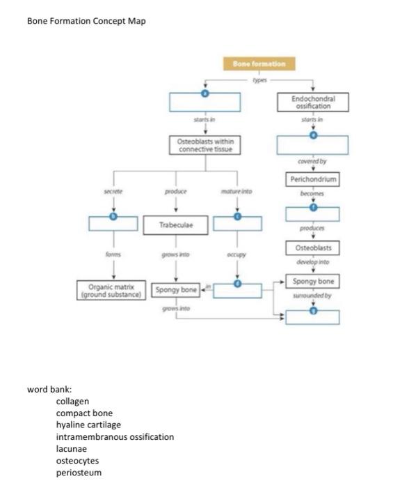
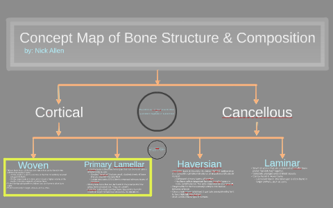
As new cartilage grows, old cartilages are converted by osteoblasts to bone cells.Intramembranous ossification and endochondral ossification.Portion between the epiphysis (shaft) medullary cavity:
This process continues until bone length increases.Intramembranous ossification occurs in flat bones like the skull and begins in utero, continuing into adolescence through mesenchymal.
Cartilage, an important connective tissue, provides structural support to other body tissues, and serves as a cushion against impacts throughout the body.Promotes reabsorption of calcium from urine.
Last update images today Concept Map Bone Formation And Growth
 Oregon Lands WR Moore, No. 3 Overall Recruit
DIJON, France -- Dutch champion Dylan Groenewegen won the sixth stage of the Tour de France after a mass sprint that was decided in a photo finish on Thursday.
Tadej Pogacar kept the yellow jersey after a nervous day on the bike amid crosswinds.
Groenewegen earned a sixth career stage victory at cycling's biggest race ahead of Biniam Girmay, the Stage 3 winner, and Fernando Gaviria, the Stage 3 runner-up. Jasper Philipsen was initially the runner-up for a second straight stage but was relegated for an irregular sprint.
"I actually don't know what happened but I was first," Groenewegen said.
There was no change among the overall leaders, with Pogacar staying 45 seconds ahead of Remco Evenepoel. Two-time defending champion Jonas Vingegaard remained in third place, 50 seconds off the pace.
After Matthieu Van der Poel opened the sprint in the city of Dijon with an excellent lead-out for his teammate Philipsen, Groenewegen timed his effort perfectly and used his great power to prevail by just a few inches.
The fight between the main contenders for the yellow jersey is expected to resume on Friday during the race's first time trial. The 25-kilometer (16-mile) race against the clock features a climb with an average gradient of 6.1% that will put riders to the test in the final section.
Thursday's relatively short stage of 163.5 kilometers (102 miles) started from Macon, taking the peloton through the Burgundy vineyards. Early into the stage, riders rode past a giant drawing of France striker Antoine Griezmann, who was born in Macon.
Jonas Abrahamsen ignited the first move of the day to claim points in the classification for the polka-dot jersey of best climber, at the top of a short climb and went on a breakaway with Axel Zingle. The duo was caught soon after.
On long sections of roads exposed to wind, the peloton rode at a steady pace, with riders careful not to get caught in a split. About 85 kilometers (53 miles) from the finish, Vingegaard's teammates Wout van Aert and Christophe Laporte moved to the front to speed up the pace and harden the race.
The peloton lined out and split in two but all the main contenders managed to stay in the first group, although Pogacar found himself isolated. It was just a scare for the UAE Team Emirates leader as the second group with his teammates managed to bridge the gap in the end.
The sprinters' teams took control with four kilometers left as the fastest men of the peloton got ready for their final, brutal effort. Groenewegen was not immediately sure he won and waited to be 100% sure before he let his joy explode with staff members of the Team Jayco AlUla.
"It was so close I couldn't celebrate on the finish line," Groenewegen said. "In the end, we grabbed it."